Doctoral Research · University of Delaware · 2019–2024
Preclinical Models · Therapeutic Evaluation · Disease Modeling
Preclinical Evaluation of Therapeutic Strategies in Age- and Treatment-Associated Tissue Degeneration
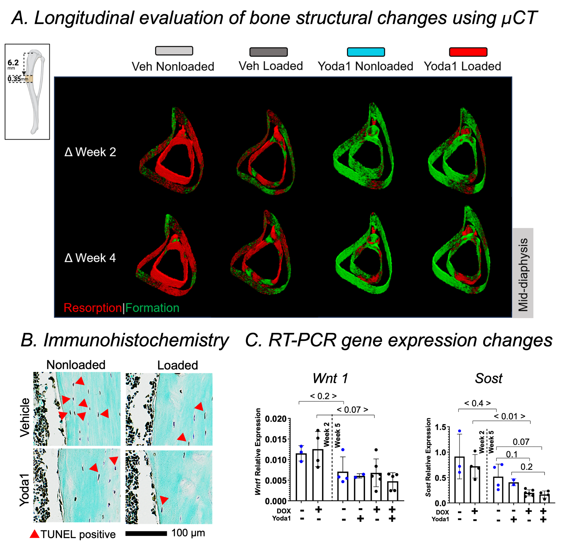
This project investigated whether mechanobiology-based interventions could mitigate
tissue deterioration associated with aging and systemic drug treatment. Using mature
mice exposed to doxorubicin, I evaluated pharmacological and mechanical interventions
using a multidimensional preclinical workflow.
The study integrated in vivo treatment, micro-CT, quantitative histology, cellular and
molecular assays, and biomechanical testing to connect therapeutic intervention with
structural, biological, and functional outcomes.
This work demonstrates my ability to design disease models, evaluate therapeutic efficacy,
and integrate multiple preclinical endpoints to characterize treatment response.
Related publication
Mitigating Aging and Doxorubicin Induced Bone Loss in Mature Mice via Mechanobiology Based Treatments
Murtaza Wasi et al. · Bone · 2024
Preclinical Models
Therapeutic Evaluation
Drug Treatment
Aging
Disease Modeling
Biomarkers
Biomechanics
Doctoral Research · University of Delaware · 2019–2024
Oncology · Disease Modeling · Quantitative Phenotyping
Therapeutic Evaluation in an Oncology Disease Model
I developed and applied a preclinical metastatic disease model to investigate how
pharmacological and mechanical interventions influence tissue integrity in the presence
of tumor-associated degeneration.
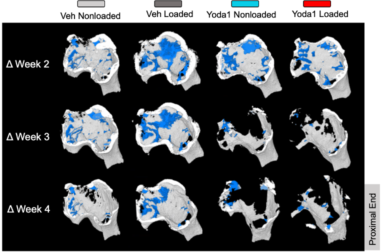
Using aged mice bearing metastatic breast cancer, I evaluated Piezo1 activation with
Yoda1 and controlled mechanical loading using spatially resolved micro-CT and biological
analyses. The study enabled assessment of regional treatment response within a complex
disease environment.
This work demonstrates experience in oncology models, pharmacological intervention,
quantitative phenotyping, and therapeutic efficacy assessment.
Related publication
Different effects of moderate tibial loading and Yoda1 on breast cancer-induced osteolysis in aged mice
Murtaza Wasi et al. · Bone · 2025
Oncology
Metastatic Disease Model
Pharmacological Intervention
Therapeutic Efficacy
Quantitative Phenotyping
Micro-CT
Spatial Analysis
Postdoctoral Research · Cornell University · Sept 2024–Aug 2026
3D Imaging · Assay Development · Computational Analysis
3D Tissue Imaging and Quantitative Image Analysis Platform Development
I develop and optimize end-to-end workflows for three-dimensional visualization and
quantitative characterization of complex biological tissues. My work spans tissue
preparation and assay optimization through high-resolution image acquisition and
computational analysis.
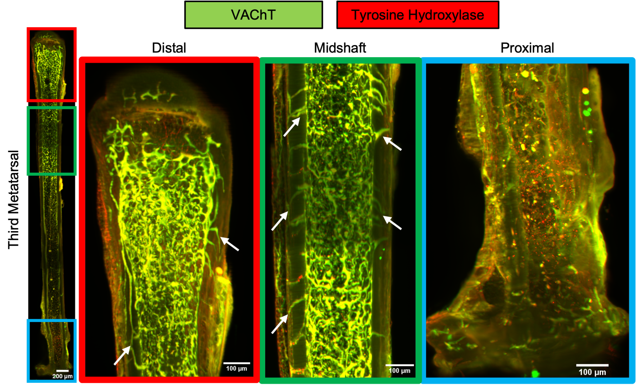
Using whole-bone innervation as a model system, I optimized tissue clearing,
immunostaining, confocal and light-sheet microscopy, and developed custom Python
workflows for segmentation, skeletonization, graph-based analysis, and morphometric
quantification of complex 3D networks.
This work demonstrates the ability to develop new imaging assays and convert
high-dimensional imaging data into reproducible quantitative biological endpoints.
Related publication
Cholinergic regulation of osteocyte mechanobiology: A paradigm for bone adaptation
Macy Mora-Antoinette, Andrea Garcia-Ortiz, Mariam Obaji, Alexander Saffari, Melia D. Matthews, Murtaza Wasi, Karl J. Lewis · Science Advances · 2025 · 11(34): eads9720 · DOI: 10.1126/sciadv.ads9720
Imaging Assay Development
Light-Sheet Microscopy
3D Imaging
Computer Vision
Python
Image Analysis
Method Development
Postdoctoral Research · Cornell University · Sept 2024–Aug 2026
In Vivo Assay Development · Pharmacology · Therapeutic Response
In Vivo Functional Assay Development for Pharmacological and Therapeutic Response
I develop in vivo functional assays to quantify cellular responses to mechanical and
pharmacological perturbations in living tissue. By integrating controlled stimulation,
localized drug delivery, intravital two-photon microscopy, and real-time calcium imaging,
I can directly measure changes in cellular function following targeted pharmacological modulation.
I have applied this platform to investigate cholinergic signaling and to characterize
functional changes following short- and long-term SOST-mAb treatment. This work supports
target validation, pharmacodynamic assessment, longitudinal drug-response phenotyping,
and mechanism-of-action studies in vivo.
Model system: osteocyte mechanosensitivity and skeletal adaptation.
In Vivo Assay Development
Pharmacology
Functional Imaging
Drug Response
Disease Modeling
Target Validation
Longitudinal Studies